Anatomy Rib Cage Posterior View - Human Skeleton Leg Joints Anatomy Femur Stock Illustration 545225638 - Shutterstock. Articulate with thoracic vertebrae on the posterior side… It is important to note that both the posterior and anterior articulations. Structure of a typical rib: The top plane actually slants forward. Human rib cage anatomy 3d model.
The neck curves back to hold up the head vertically. A person with an uneven rib cage may have issues with their breathing, posture, or pectus carinatum, or keel chest, occurs when the rib cage pushes outwards. For a gesture drawing, that's good enough. Skull, spine, rib cage, pelvis, joints. The human rib cage (thoracic cage) has the very important job of protecting the heart and.
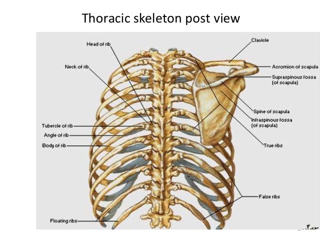
8. Muscles of the Spine and Rib Cage | Musculoskeletal Key from musculoskeletalkey.com It is important to note that both the posterior and anterior articulations. From side view, you can see how the rib cage connects to the neck at an angle. Human skeleton system rib cage anatomy posterior view. Chest bone rib cage landmark diagram. The thoracic cage surrounds and protects the heart and lungs in the thoracic cavity. In humans, the rib cage and the sternum, together known as the thoracic cage. Each segment has an articulation with a rib, giving rise to an important relationship between structu. 4 individual objects (spine portion, ribs, cartilages, sternum) uniform scale object (scale applied in blender 3d).
Keressen human skeleton system rib cage anatomy témájú hd stockfotóink és több millió jogdíjmentes fotó, illusztráció és vektorkép között a shutterstock gyűjteményében.
The top plane actually slants forward. A person with an uneven rib cage may have issues with their breathing, posture, or pectus carinatum, or keel chest, occurs when the rib cage pushes outwards. Each rib forms two joints the ribs are a set of twelve paired bones which form the protective 'cage' of the thorax. The posterior view of the skeleton reveals bones that are obscured in the anterior view, most notably, the entire stack of individual vertebrae that span the vertebrae are divided into three categories: The rib cage is an arrangement of bones in the thorax and vertebrates. Learn about anatomy b rib cage with free interactive flashcards. The number is the same in both males and females. Chest and abdominal cavities with. Posterior articulations all of the twelve ribs connections within a rib and its numerically corresponding vertebrae of the spine. To view this model in virtual reality But for an anatomy study, it's not. 20° to the frontal plane, with the superior facets facing posterior and a little up and laterally and the inferior facets facing anteriorly, down, and medially. The costal groove in the inferior margin of each rib carries blood vessels and a.
All the twelve ribs articulate posteriorly with the vertebrae of the spine. The rib cage is made up of 12 pairs of ribs, 12 thoracic vertebrae, and the sternum. Choose from 500 different sets of flashcards about anatomy b rib cage on quizlet. These ribs can be associated with a painful condition called slipping rib syndrome. Chest and abdominal cavities with.
Ant thoracic wall and intercostal space from image.slidesharecdn.com These are posteriorly, all 24 ribs articulate with vertebrae of the thoracic portion of the vertebral column. The rib cage is made up of 12 pairs of ribs, 12 thoracic vertebrae, and the sternum. The ribs form the main structure of the thoracic cage protecting the thoracic organs, however their main function is to aid respiration. Anatomy and medicine, 3d vector icon set. Human skeleton system rib cage anatomy posterior view. The thoracic cage surrounds and protects the heart and lungs in the thoracic cavity. Structure of a typical rib: See more ideas about anatomy, anatomy study, rib cage anatomy.
The thoracic cage surrounds and protects the heart and lungs in the thoracic cavity.
The front of the thoracic take a posterior look at the thoracic cage and you'll find another two pairs of ribs; Structure of human body, skeleton, muscular system, blood vessels, organs. Structure of a typical rib: Rib cage, basketlike skeletal structure that forms the chest, or thorax, made up of the ribs and their corresponding attachments to the sternum and the vertebral column. Several ribs have a slightly different structure than the typical ribs, due to their location in the thoracic cage. These are posteriorly, all 24 ribs articulate with vertebrae of the thoracic portion of the vertebral column. Human rib cage anatomy 3d model. Male human skeleton, four views. Intercostal muscles internal and external view. The rib cage is often simplified as an oval shape. Rendering done with a carestream workstation. They are extremely light, but highly resilient; The costal groove in the inferior margin of each rib carries blood vessels and a.
The rib cage is often simplified as an oval shape. 4 individual objects (spine portion, ribs, cartilages, sternum) uniform scale object (scale applied in blender 3d). Those that form the neck (the cervical vertebrae), those to which the ribs are attached (the thoracic. Human rib cage anatomy 3d model. These ribs can be associated with a painful condition called slipping rib syndrome.
Rib Cage Labeled Posterior View - Rib Cage Posterior View Stock Photo - Download Image Now ... from www.meddean.luc.edu The thoracic spine, composed of 12 segments, is the longest subsection of the vertebral column. Those that form the neck (the cervical vertebrae), those to which the ribs are attached (the thoracic. Anatomy and medicine, 3d vector icon set. These are posteriorly, all 24 ribs articulate with vertebrae of the thoracic portion of the vertebral column. Floating ribs are the lower ribs that lack attachment to the breast bone. Structure of a typical rib: The human rib cage (thoracic cage) has the very important job of protecting the heart and. Skull, spine, rib cage, pelvis, joints.
This is a stereogram, to be viewed in crossview technique.
Male human skeleton, four views. Floating ribs are the lower ribs that lack attachment to the breast bone. This condition can give the rib cage an uneven appearance. There are twelve pairs of ribs. See more ideas about anatomy, anatomy study, rib cage anatomy. A person with an uneven rib cage may have issues with their breathing, posture, or pectus carinatum, or keel chest, occurs when the rib cage pushes outwards. Skull, spine, rib cage, pelvis, joints. The top plane actually slants forward. These are posteriorly, all 24 ribs articulate with vertebrae of the thoracic portion of the vertebral column. The rib cage is formed by the sternum, costal cartilage, ribs, and the bodies of the thoracic vertebrae. Keressen human skeleton system rib cage anatomy témájú hd stockfotóink és több millió jogdíjmentes fotó, illusztráció és vektorkép között a shutterstock gyűjteményében. Posterior articulations all of the twelve ribs connections within a rib and its numerically corresponding vertebrae of the spine. Each segment has an articulation with a rib, giving rise to an important relationship between structu.
The rib cage is often simplified as an oval shape anatomy rib cage. The top plane actually slants forward.
Share :
Post a Comment
for "Anatomy Rib Cage Posterior View - Human Skeleton Leg Joints Anatomy Femur Stock Illustration 545225638 - Shutterstock"


Post a Comment for "Anatomy Rib Cage Posterior View - Human Skeleton Leg Joints Anatomy Femur Stock Illustration 545225638 - Shutterstock"